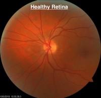

Diabetic retinopathy results from damage to the blood vessels within the retina, the tissue that lines the back of the eye. The retina is where the light rays that enter the eye are focused. This information is transported via the optic nerve to the back of the brain where it is processed into the pictures we see. With diabetic retinopathy, the blood vessels within the retina leak fluid and bleed. This can affect a person’s ability to see clearly.
4 Stages of Diabetic Retinopathy:
• Mild nonproliferative retinopathy: The blood vessels in the retina develop small areas of balloon-like swelling called microaneurysms. There are likely no visual symptoms at this stage.
• Moderate nonproliferative retinopathy: As the disease process progresses, the blood vessels continue to swell. There is increased bleeding and leaking of fluid from the vessels that may result in swelling within the retinal tissue. This is the stage when visual symptoms often develop due to the swelling of the retinal tissue.
• Severe nonproliferative retinopathy: At this stage, the blood flow within some of the vessels becomes blocks. This results in a lack of blood supply to areas of the retina. These areas will then secrete growth factors that signal the retina to grow new blood vessels. Visual symptoms will likely worsen.
• Proliferative diabetic retinopathy: The growth factors secreted by the retina have now caused the development of new blood vessels at this stage. These new blood vessels grow along the inside surface of the retina and into the jelly that fills the back 2/3 of the eye, the vitreous. These new blood vessels are fragile, making them more likely to bleed and leak fluid. Scar tissue may form. This scar tissue can contract and cause a retinal detachment, which can lead to permanent vision loss.
How is Diabetic Retinopathy Detected?
A comprehensive, dilated eye exam allows the doctor to exam the retina to look for signs of diabetic retinopathy. The doctor will look for any changes in the blood vessels within the retinal tissue. Leaking blood vessels and fatty deposits can be viewed through the dilated pupil. Swelling can be noticed with the use of a special magnifying lens and a test called an OCT, optical coherence tomography. The OCT will provide detailed, cross-sectional images of 10 layers of the retina.
Our next blog will discuss how diabetic retinopathy is treated. If you need a dilated eye exam due to diabetes, call Summit Eye Center at (816) 246-2111 or email